How Laser Diffraction Works: Simple Explanation
Have you ever wondered how scientists measure the size of tiny particles, from nanoparticles in sunscreen to metal powders used in 3D printing? Laser diffraction is one of the most powerful and widely used methods for particle size analysis. This fast, non-invasive technique can determine particle size distributions across an extraordinary range, from 10 nanometers to over 3 millimeters, all in a matter of seconds. Whether you’re testing pharmaceuticals, food emulsions, or industrial powders, laser diffraction delivers rapid, reproducible results by analyzing how light bends and scatters when it hits particles.
The core principle is straightforward: smaller particles scatter light at wider angles, while larger particles scatter light narrowly forward. By capturing the angular pattern of this scattered light and applying sophisticated optical theories, instruments convert raw data into precise particle size distributions. In this guide, we’ll break down the physics, instrumentation, data processing, and real-world applications of laser diffraction so you understand not just what it measures, but how it works under the hood.
Core Physics: Why Light Scattering Reveals Particle Size
When a laser beam hits particles, it doesn’t simply bounce off. Multiple optical phenomena occur simultaneously, and the resulting scattering pattern holds the key to determining particle size. Understanding these interactions is essential for interpreting results correctly.
How Diffraction Dominates for Large Particles
For particles larger than approximately 20 micrometers, edge diffraction becomes the primary mechanism. As light waves pass around a particle, they bend and interfere, creating a distinct angular pattern. This follows a predictable principle: the smaller the particle, the wider the diffraction cone. Large particles produce tightly focused forward scattering, while submicron particles spread light across wide angles.
This behavior is described by Fraunhofer diffraction theory, which assumes particles are opaque and much larger than the laser wavelength. It’s computationally fast and useful for coarse powders, but limited when dealing with transparent or nanoscale materials.
Why Refraction Matters for Small Particles
Below approximately 20 micrometers, refraction becomes critical. When light enters a transparent particle, it changes direction due to differences in refractive index between the particle and surrounding medium. The light path continues with internal reflections and interference effects before exiting.
These complex interactions require a more rigorous model called Mie scattering theory. Unlike Fraunhofer, Mie accounts for refraction, reflection, absorption, and interference within the particle. Mie theory applies across the full measurement range, from 10 nanometers to 3 millimeters, but demands accurate input of the complex refractive index. Even small errors in refractive index or absorption can shift results significantly, especially for fine particles.
Rayleigh Scattering at the Nanoscale
For particles far smaller than the wavelength, Rayleigh scattering dominates. In this regime, scattering intensity increases with the sixth power of diameter, and light scatters almost equally in all directions. This makes detection extremely challenging because tiny drops in size cause massive signal loss. While Rayleigh theory helps explain detection limits, modern instruments rely on enhanced optics and dual-wavelength lasers to overcome these challenges.
Key Instrument Components and Their Functions
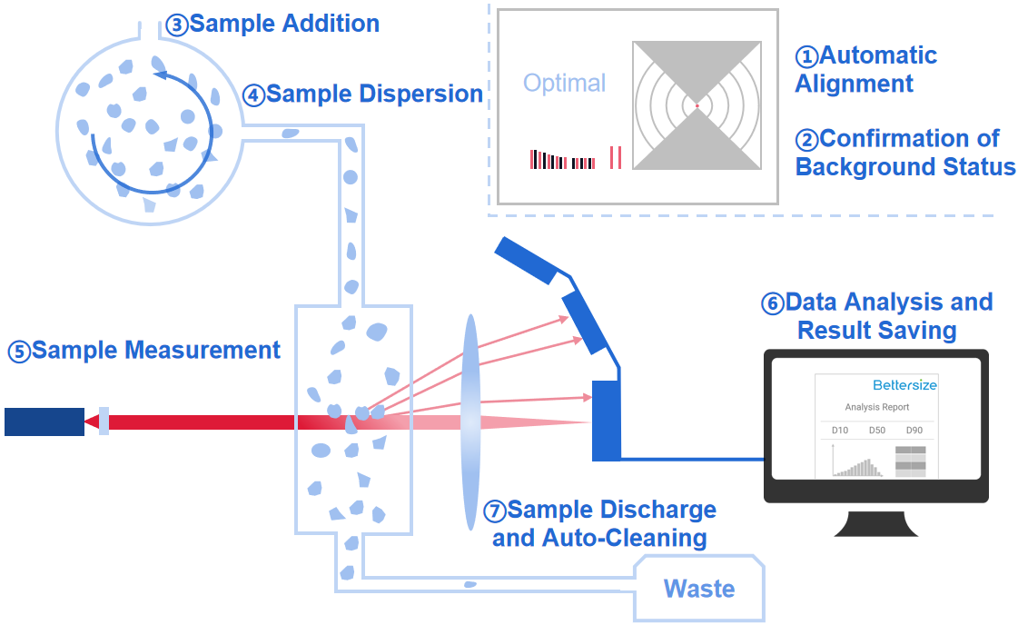
Modern laser diffraction analyzers are engineered for precision, speed, and wide dynamic range. Each component plays a critical role in capturing accurate scattering patterns.
Light Source and Wavelength Selection
The choice of laser wavelength directly impacts sensitivity to small particles. Red lasers at 633 to 650 nanometers are standard for particles larger than 1 micrometer. Blue lasers at 405 to 450 nanometers boost sensitivity below 1 micrometer because scattering intensity increases with the inverse fourth power of wavelength. Advanced systems use dual-wavelength setups to extend the measurable range down to 30 nanometers without changing optics.
Beam Conditioning for Precision Illumination
Raw laser beams aren’t uniform and require shaping. Beam expanders, spatial filters, and collimating lenses ensure a clean, parallel beam, consistent illumination across the measurement zone, and minimal divergence. This conditioning is essential for accurate angular measurements, especially in Fourier optics systems.
Sample Dispersion Methods
Particles must be fully dispersed to avoid false readings. Two main methods exist.
Wet dispersion is used for liquids and emulsions. The process involves stirring to suspend particles, ultrasonication to break up agglomerates, and controlled pump speed to prevent re-agglomeration. This method is ideal for pharmaceutical suspensions or food emulsions.
Dry dispersion uses compressed air through a Venturi nozzle to disperse powders via particle-particle and particle-wall collisions. This method suits moisture-sensitive materials, toners, and inhaler formulations. Critical to optimize air pressure because too low causes poor dispersion and too high may fragment particles.
The Measurement Zone and Optical Configuration
The measurement zone is where light meets particles. Two optical configurations exist.
Fourier optics uses a parallel beam, allowing particles to pass anywhere in the beam cross-section while angular scattering remains consistent. This delivers sharper, more accurate patterns and is preferred in high-end instruments.
Reverse Fourier optics uses a convergent beam requiring precise particle positioning in the focal region. There’s risk of blurred patterns due to positional variation, but this allows smaller detector sizes and is used in cost-effective systems.
Detector Array Design
A multi-element photodetector captures scattered light from 0 degrees to over 175 degrees. Key features include a central detector for forward scattering from large particles, annular rings for side and back-angle scattering from small particles, and a central aperture that lets the unscattered beam pass without saturation. High angular resolution allows detection of subtle differences, which is critical for resolving multimodal distributions.
Understanding the Scattering Theories: Fraunhofer vs Mie
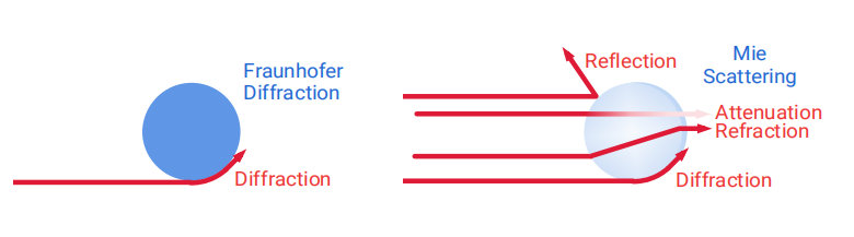
Choosing the right theoretical model affects accuracy, especially for small or transparent particles.
When Fraunhofer Theory Works Best
Fraunhofer theory is best for particles larger than 10 to 20 micrometers and opaque materials with unknown refractive index. The advantages include no need for optical properties and computationally efficient calculations. However, the limitations are significant: inaccurate for submicron or transparent particles and ignores refraction and internal scattering. This theory is still widely used in industrial quality control for coarse powders.
Why Mie Theory Is the Gold Standard
Mie theory applies to the full range from 10 nanometers to 3 millimeters, provided the complex refractive index is known. It accounts for diffraction, refraction, reflection, and absorption, matching real-world scattering behavior. This makes it essential for particles smaller than 20 micrometers. The requirement is accurate refractive index values, which are often obtained from literature or optimized via fit quality metrics.
Rayleigh Theory’s Limited Practical Use
Rayleigh theory is only valid for particles much smaller than the wavelength, such as less than 40 nanometers. It’s rarely used directly in software because isotropic scattering makes it hard to resolve angles, the signal is very weak, and noise dominates. This theory mainly helps understand lower detection limits rather than providing practical sizing data.
Data Processing: From Raw Signals to Size Distributions
Raw detector output is just intensity versus angle. Converting this into meaningful particle size data requires advanced mathematical inversion.
The Inversion Problem Explained
The fundamental equation behind laser diffraction is I = C × N, where I represents measured intensity at each angle, C represents the theoretical scattering matrix based on Mie or Fraunhofer theory, and N represents the volume distribution across size classes. This is an ill-posed inverse problem because many size distributions could theoretically produce the same pattern. The goal is finding the most physically plausible N that minimizes error.
Algorithms Used for Data Processing
Common solution methods include non-negative least squares, which ensures all volume fractions are positive, regularization techniques that prevent overfitting and stabilize results, and iterative refinement that improves fit quality for complex samples. Software tools often display fit quality metrics, and values close to zero indicate excellent agreement between measured and calculated patterns.
Understanding Volume-Weighted Output
Laser diffraction reports volume-based size distributions, meaning a single 10 micrometer particle contributes 1,000 times more volume than a 1 micrometer particle. Larger particles dominate the distribution. Key metrics include D10, where 10 percent of volume is below this size, D50 or median where half the volume is above and half below, and D90 where 90 percent of volume is below this size. The span, calculated as D90 minus D10 divided by D50, indicates the width of distribution.
Detection Limits and Measurement Range
Laser diffraction offers one of the broadest ranges of any sizing technique.
Size Range Capabilities
The theoretical lower limit is 10 nanometers, while the practical lower limit is 30 nanometers with blue laser and enhanced detection. The upper limit reaches up to 4 millimeters, giving a dynamic range up to 1 to 400,000. No lens changes or manual adjustments are needed across the full range.
Technologies Enhancing Submicron Detection
Three technologies boost nanoparticle sensitivity. Dual-wavelength lasers use red and blue to increase angular contrast. Extended-angle detection up to 175 degrees captures wide-angle scattering from small particles. PIDS, or polarization intensity differential scattering, uses polarized light to detect size-dependent phase shifts in small particles. Systems equipped with these technologies improve resolution below 500 nanometers.
Sample Preparation Best Practices
Even the best instrument gives bad data with poor sample preparation.
Optimizing Obscuration Levels
Ideal obscuration is 10 to 20 percent, corresponding to 80 to 90 percent transmission. Too high causes light scattering off multiple particles, distorted patterns, and artificially large size values. Too low reduces the signal-to-noise ratio.
Following Standard Operating Procedures
Standard operating procedures should define dispersant type, sonication time, pump speed or air pressure, measurement duration, and refractive index settings. Automation ensures consistency, which is critical in regulated industries like pharmaceuticals.
Avoiding Common Errors
Key errors to avoid include agglomeration that causes artificial shifts to larger sizes, incorrect refractive index that skews results especially below 20 micrometers, multiple scattering from excessive concentration, poor dispersion that leads to inaccurate D90 values, and multimodal samples that may require advanced algorithms.
Common Limitations and Assumptions You Must Know
Laser diffraction is powerful, but it makes assumptions that affect interpretation.
The Spherical Particle Model
The system assumes every particle is a perfect sphere. Real-world implications include fibers, platelets, or rods being reported as equivalent spherical diameter, which reflects light-scattering behavior rather than true geometry. A 10 micrometer fiber may report as 6 micrometer equivalent spherical diameter. This is why laser diffraction results often differ from microscopy.
Other Key Limitations
Agglomeration causes artificial shifts to larger sizes. Incorrect refractive index skews D-values, particularly for particles below 20 micrometers. Multiple scattering creates broadened or bimodal artifacts. Poor dispersion leads to inaccurate D90 and overestimated fines. Always validate with orthogonal methods like dynamic light scattering or electron microscopy if results seem off.
Industrial Applications and Use Cases
Laser diffraction is the most widely adopted particle sizing method globally, supported by ISO, USP, and ASTM standards.
Pharmaceutical Applications
The pharmaceutical industry uses laser diffraction for API particle sizing that affects dissolution rate, dry powder inhalers where D50 impacts lung deposition, and emulsion stability in injectables. It complies with USP 429 and ISO 13320:2020.
Food and Cosmetic Applications
Examples include milk emulsions for controlling mouthfeel and shelf life, chocolate powders to ensure flow and mixing, and sunscreens to verify nanoparticle dispersion.
Advanced Manufacturing
Carbon black and quantum dots require confirmation of nanoscale distribution. 3D printing metal powders need verification of flowability and packing density. Environmental applications include soil texture analysis for clay, silt, and sand, aerosol monitoring in air quality studies, and sediment transport modeling.
Standards and Quality Assurance
Unlike other instruments, laser diffraction doesn’t require routine calibration because it’s based on first-principle physics.
Key Standards to Know
ISO 13320:2020 defines instrument validation and measurement procedures. USP 429 provides regulatory compliance in pharmaceuticals. ISO 9276 guides reporting size distributions.
Performance Verification Methods
Use NIST-traceable standards like polystyrene latex spheres and silica suspensions. Check repeatability where relative standard deviation should be less than 1 to 2 percent, resolution for ability to distinguish close peaks, and background stability. Monitor fit quality parameters and obscuration in every run.
Frequently Asked Questions About Laser Diffraction
What is the size range for laser diffraction particle analysis?
Laser diffraction can measure particles from approximately 10 nanometers to over 3 millimeters in a single measurement. Practical limits typically range from 30 nanometers to 1 millimeter depending on the instrument configuration.
Why do laser diffraction results differ from microscopy?
Laser diffraction assumes spherical particles and reports equivalent spherical diameter based on light-scattering behavior. Microscopy measures actual geometric dimensions. The different definitions and volume weighting versus number weighting explain the discrepancies.
Do I need to calibrate my laser diffraction analyzer?
No routine calibration is required because laser diffraction is based on first-principle physics. Periodic verification with NIST-traceable reference materials ensures the instrument is performing correctly.
Which scattering theory should I use for my samples?
Use Mie theory for all samples, especially those below 20 micrometers or with transparent particles. Fraunhofer theory is acceptable only for opaque particles larger than 20 micrometers when optical properties are unknown.
How does sample preparation affect laser diffraction results?
Poor dispersion causes agglomeration, leading to artificially large particle sizes. Multiple scattering from excessive concentration distorts patterns. Following standard operating procedures for dispersant selection, sonication, and concentration is essential for accurate results.
Can laser diffraction measure non-spherical particles?
Laser diffraction measures equivalent spherical diameter, which represents the diameter of a sphere that would scatter light identically to the actual particle. Non-spherical particles will report as different sizes depending on their orientation and shape.
Key Takeaways for Understanding Laser Diffraction
Laser diffraction works by measuring how particles scatter light at different angles and using physics-based models to convert that pattern into a volume-weighted size distribution. It combines speed, wide range, and high reproducibility, making it the go-to method for particle sizing in science and industry.
The technique relies on fundamental scattering physics where smaller particles scatter light at wider angles and larger particles scatter narrowly forward. Modern instruments use dual-wavelength lasers, Fourier optics, and sophisticated inversion algorithms to achieve measurement ranges from 30 nanometers to 4 millimeters.
While laser diffraction assumes spherical particles and requires careful setup including proper dispersion and accurate refractive index input, its strengths include no calibration needed, single-measurement range across multiple orders of magnitude, and compliance with global standards. Whether you’re developing a new drug, optimizing a food product, or qualifying 3D printing powders, understanding how laser diffraction works empowers you to get accurate, meaningful results every time.